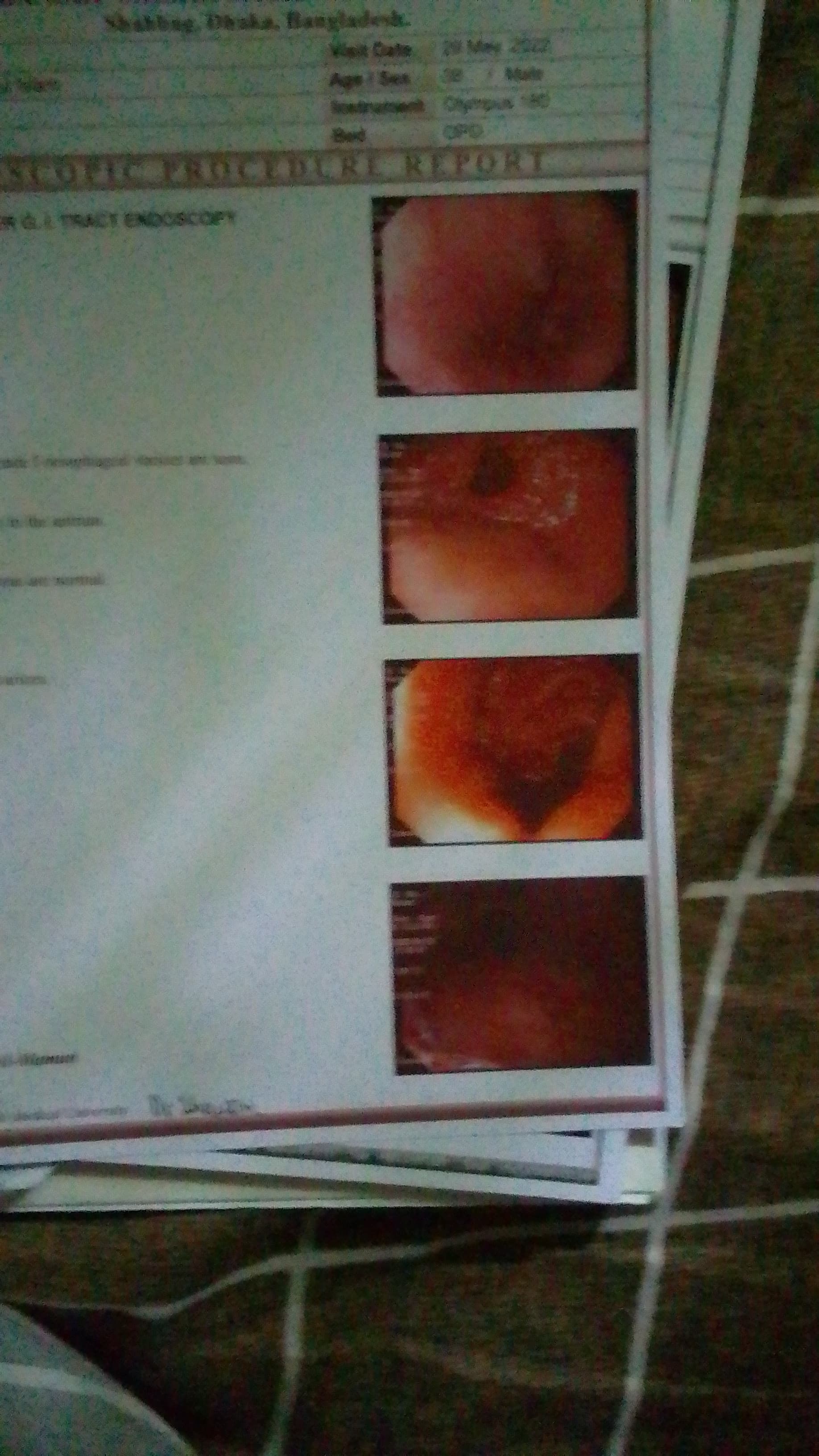
Assalamualaikum, sir my Upper GI track endoscopy , Oesophagus: multiple columns of grade I (1) oesophageal varices are seen ,
Stomach : multiple erosion seen in the antrum.
Duodenum: bulb and post bulb areas are normal,
Comments: grade I (1) oesophageal varices .
Hepatitis b DNA un detected.
Alt ( SGPT : 25 ul
HBeAg : n
egative.
Bilirubin: 0.70 mg/dl
VDRL : non reactive
What is my condition,
Please your advice
Dear @Saiful_Sobahan,
Welcome to the forum. Oesophageal varices mean that the blood vessels around your neck and stomach are bigger than usual (sort of like a balloon that is too full).
The most common cause of varices is if you have liver injury: if your liver is hurt, then it is harder to pump blood through it and you have blood high pressure in this system. Because the veins in your liver are connected to the ones in your neck, it puts pressure on these veins.
The blood results here do not show whether you have chronic HBV or not.
It is important to consult with your doctor about how to take care so the varices and stomach erosion do not get worse because these conditions can be dangerous.
1 Like
Dear Saiful,
I see your HBV DNA is undetectable and you HBeAg test is negative with normal ALT and bilirubin. Are you currently receiving treatment for HBV infection?
No, still not taken any medicine.
Dear Saiful,
Varices are a sign of portal hypertension (a sign of advanced liver disease) and it seems that your doctor has done some preliminary testing to see if this could be due to HBV infection. If you are not receiving antiviral treatment for HBV, your results indicate that you do not have an active HBV infection so the cause for your portal hypertension appears to be something else.
I agree with Thomas that you should not wait to deal with this funding.
Unfortunately, an upper GI evaluation does not tell us much about what is going on in the liver. You should have a liver ultrasound done. Also, have you been tested for HCV infection?
Best of luck.
2 Likes
I agree with @availlant, and just want to ask as well have you had a HBsAg and anti-HBs test? These are essential to knowing if you have a chronic HBV infection still and they don’t seem to be in the results you have posted.
Thomas
1 Like
Sir : test results: AFP : 10.30ng / ml up to 13.6 Ng/ml
Pt result 13.20 seconds ref 12.00 -16.00 second
Creatinine serum : 1.43 mg/dl ref : 0.6-1.3mg /dl
Albumin : results : 49gm/L ref 32_48 gm/L
Dear Saiful,
You liver function appears normal and AFP is within the normal range.
I would talk to your doctor about a liver ultrasound.
Best regards,
CT scan of hbs with contrast:
Multiple axial non- contrast and contrast CT scan were permed .
Findings :
Liver is normal in size (14.2 cm ) . No focal lesion is noted .
Portal vein measures about 11.6 mm.
Intra hepatic biliary tress are not dilated .
Gallbladder is normal in size .wall thickness with normal limit. No radio opoque calculas is noted within it .
Spleen is normal in size,(9cm).no focal lesion is noted.
Pancreas is normal in.shape . position with uniform with tissue attenuation. No focal lesion is seen .MPD is not dilated,
Both kidneys are normal in size.shape and axial orientation with normal excretion of contrast media. Both ureters Re not dilated.
Bowel loops is the fields of view reveal no abnormality.
No ascites is noted .
Multiple enlarged and subcentrimetric lymphnodes are noted at periportal, peripatetic and left para aortic regions, largest one measuring about 1.5 cm in peripancreatic region .after IV contrast administration .no abnormal enchancement is noted .
Impression : abdominal lymphadenopathy.
Please advice
Hi @Saiful_Sobahan,
I think this is beyond the expertise of this community as there doesn’t seem to be anything to do with Hep B in this case. I personally am not confident in providing any input on these CT results at all.
The best thing to do in this instance is talk to your doctor about the results and what they mean in your specific case.
Thomas
1 Like